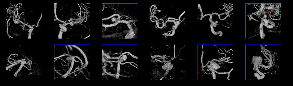
Description:
These models of cerebral aneurysms were constructed from 3D rotational angiography
images. Physiologic flow conditions were not available for these
patients, therefore flow measurements in the arteries of the circle of
Willis obtained on a normal subject were used. These measurements were
performed using phase-contrast MR.
The following figure shows volume renderings of the 3DRA images.
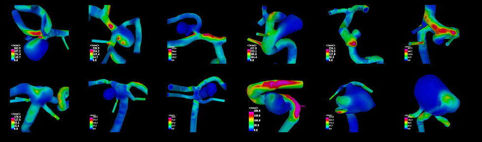
The corresponding anatomical models are shown below:

Numerical calculations of the flow of blood in these models were
performed using a finite element method.
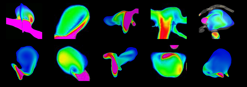
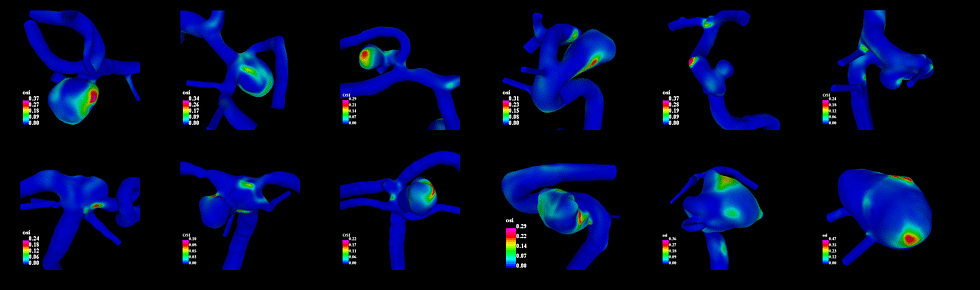
Visualizations of the mean wall shear stress are shown below:
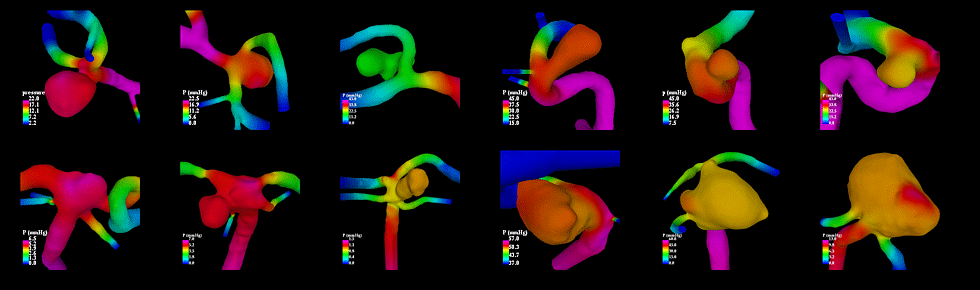
The following figure shows the systolic pressure distribution:
Oscillatory shear index are shown below:
And finally, visualizations of the blood flow pattern are presented
below. These images show that there are different types of flow
patterns. The flow in some aneurysms is stable and has only one main
vortex, while in others there is a more complex vortex structure that
remains stable during the cardiac cycle, and yet in others the flow
pattern is unstable and very dynamic throghout the cardiac cycle.