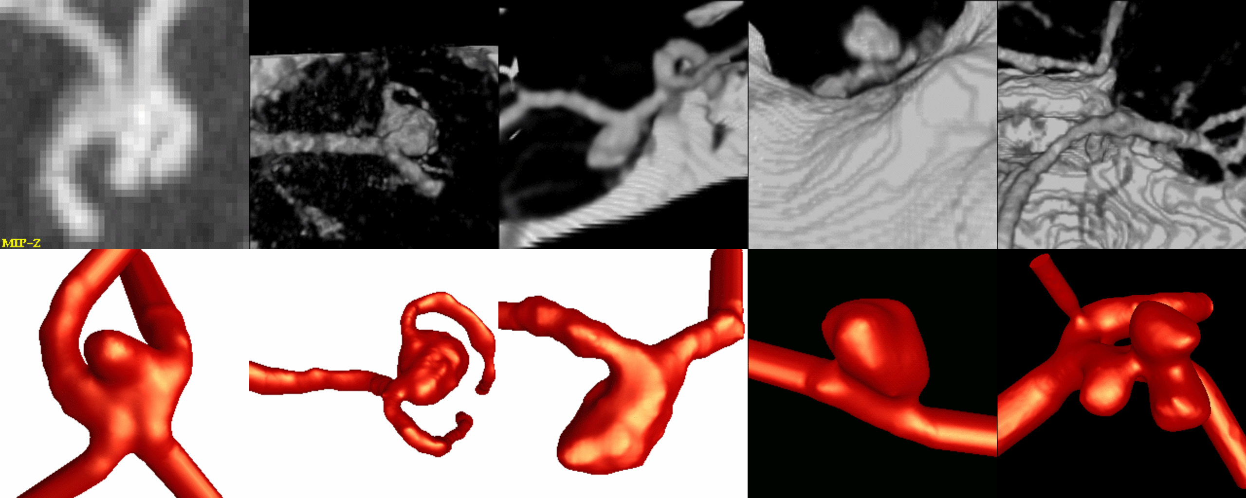
Description:
These models of cerebral aneurysms were constructed from CTA
images. Physiologic flow conditions were not available for these
patients, therefore flow measurements in the arteries of the circle of
Willis obtained on a normal subject were used. These measurements were
performed using phase-contrast MR.
The following figure shows volume renderings of the CTA images and the corresponding geometry reconstructions.
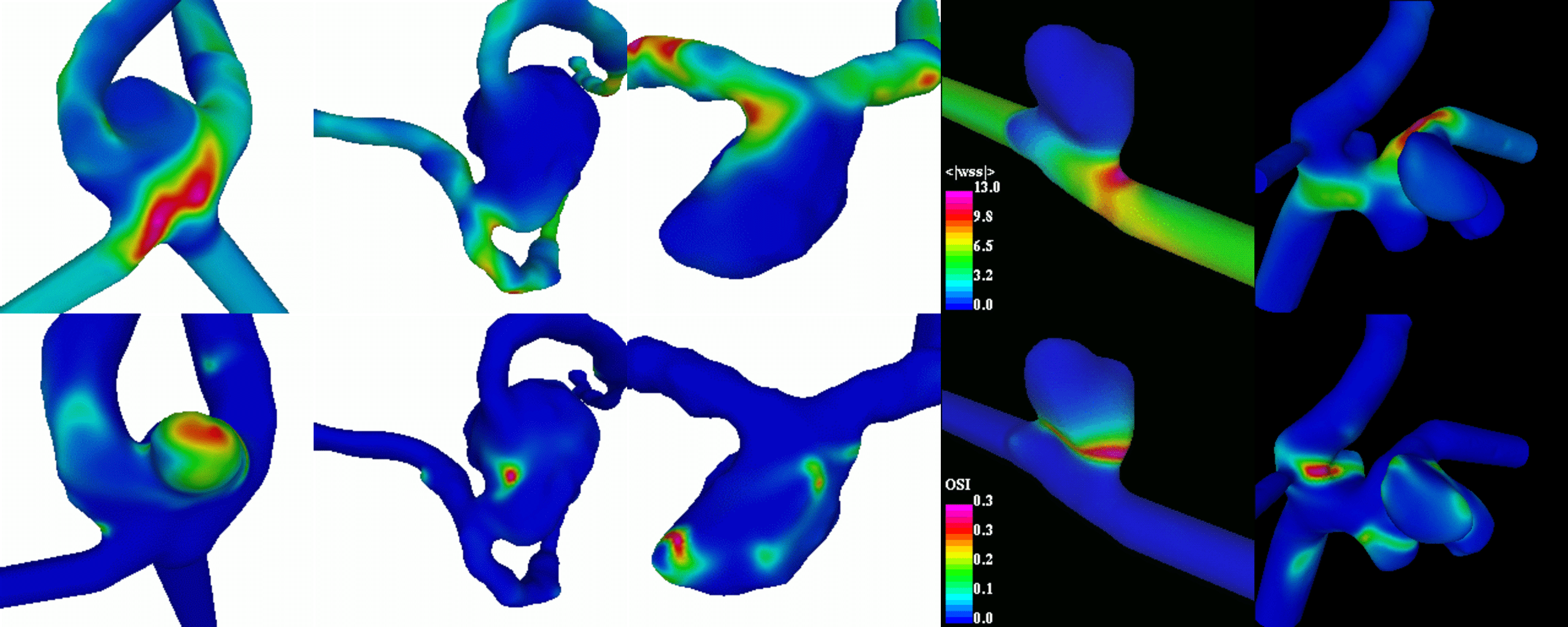
Numerical calculations of the flow of blood in these models were
performed using a finite element method. Visualizations of the mean
wall shear stress and oscillatory shear index are shown below. These
images show that the distribution of these hemodynamic quantities may
be different between aneurysms, even between aneurysms of the same kind.