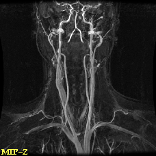

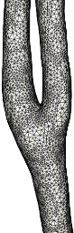
Maximum intensity projection (MIP) of an MRA and the reconstructed
model of the right carotid artery
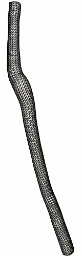

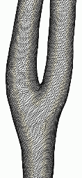
Models of the internal, external and common carotid arteries, the merged
model and the finite element grid
Realistic arterial models are reconstructed from anatomical images using
tubular deformable models. First the vessel axis of each arterial branch
is interactively selected on cross-sectional views (or obtained from skeletonization
algorithms). Then a tubular deformable model along each vessel is constructed
and allowed to deform due to forces from the image intensity gradient.
The resulting triangulations are then merged into a water-tight surface
using an adaptive voxelization technique. This triangulated surface is
used as
a support surface or geometric definition of the computational domain
for finite element grid generation. Unstructured grids composed of tetrahedral
elements are generated using an advancing front method. Element sizes can
be automatically specified from the surface curvature using adaptive background
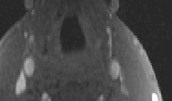
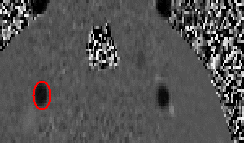
grids. A model constructed from magnetic resonance angiography (MRA) images
is shown below.
Maximum intensity projection (MIP) of an MRA and the reconstructed
model of the right carotid artery
Models of the internal, external and common carotid arteries, the merged
model and the finite element grid
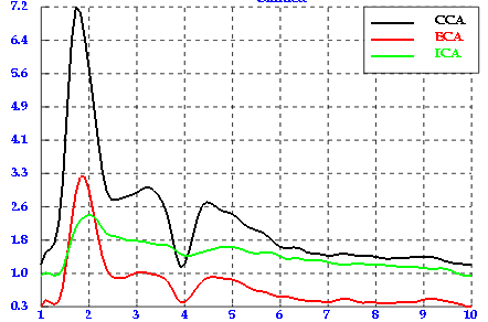
Physiologic flow conditions are derived from phase-contrast magnetic
resonance (PC-MR) measurements of blood flow velocity. Flow rate curves
along each arterial branch are obtained by integration of the velocity
profile over the cross section of the vessel. The vessel lumen is defined
manually, or segmented from the magnitude images, or automatically segmented
from the phase images using velocity cross-correllations. Pulsatile velocity
boundary conditions are typically imposed using a supperposition of Womersley
profiles for each Fourier mode.
|
 |
The methodology described above was applied for the reconstruction of numerous normal and diseased carotid arteries from different imaging modalities including contrast-enhanced magnetic resonance angiography, CT angiography and 3D rotational angiography. The pictures below show some of the reconstructed models. These images illustrate the large anatomical variability among individuals. Since the flow dynamics strongly depends on the vessel shape, subject-specific models yield greater insight into arterial hemodynamics than computational models based on idealized geometries. Furthermore, patient-specific models are required for improved diagnosis and treatment planning.
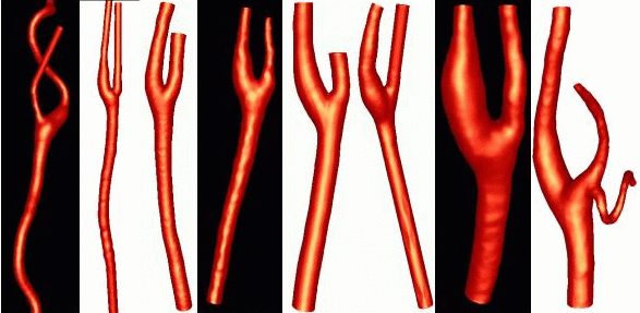
Models of normal carotid arteries
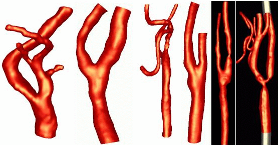
Models of athersclerotic carotid arteries