

Description:
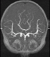
This example illustrates the blood flow calculation in a model of the circle of Willis reconstructed from contrast-enhanced MRA images, using a region growing algorithm.
The following images show some of the image processing stages.


The next images show the reconstructed arterial tree and the cutting of the final model.
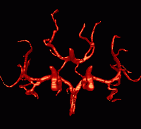
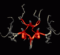
The next two images show the final geometrical model from two different view-points.
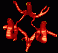
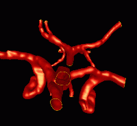
The following images show the generated finite element grid from two view-points.
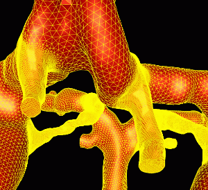
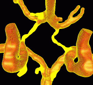
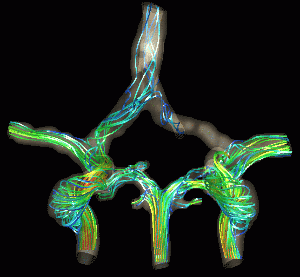
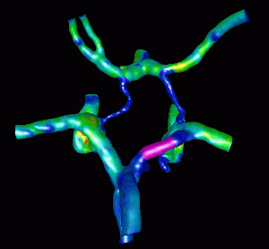
The next figures show the application of CFD modeling to surgical planning. In this case one of the cerebral arteries is temporarily occluded and the new flow redistribution is calculated. The flow patterns before and after the occlusive maneuver has been carried out are shown below.
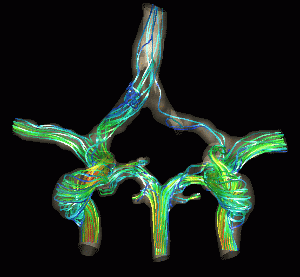
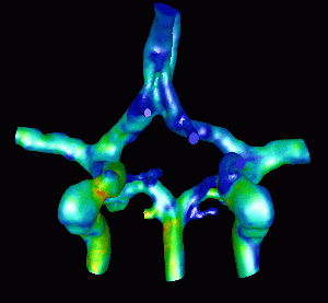
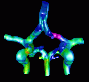
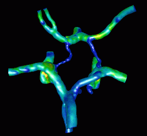
Finally the next series of images show the wall shear stress distributions before and after the occlusion from two different view-points. The magenta region represents the occluded region.
Observations: