The model was reconstructed using a tubular deformable model along each arterial branch, followed by surface merging using an adaptive voxelization technique. The mode was then smoothed and cut perpendicularly to the vessel axis in order to impose boundary conditions. An unstructured grid composed of tetrahedral elements was then generated using an advancing front method that operates directly on surface triangulations, i.e. does not require an analytical representation of the coputational domain to be meshed.
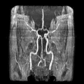
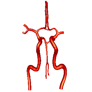
MIP of the MRA images reconstructed model
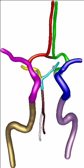
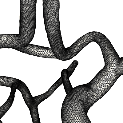
reconstructed branches finite element grid
The solution of the unsteady 3D Navier-Stokes equations for an incompressible fluid were then numerically solved using an implicit finite element formulation. Pulsatile flow boundary conditions were derived from time dependent flow rates derived from the PC-MR measurements. Visualization of hemodynamic quantities were then produced:
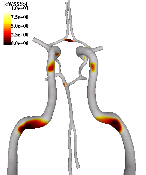
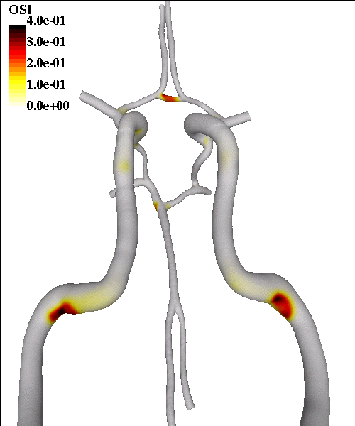
mean wall shear stress magnitude oscillatory shear index
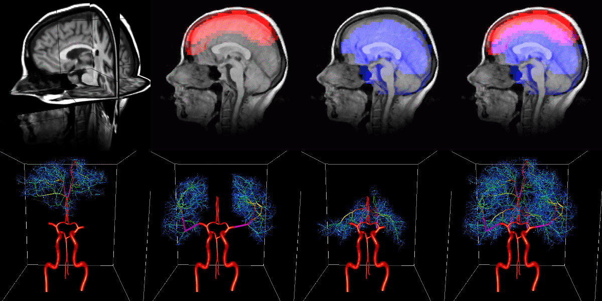
Finally, we are investigating the possibility of using arterial tree models generated from anatomical images of the brain to impose outflow boundary conditions for these 3D models of the circle of Willis. These models can also be used to estimate local tissue perfusion, by solving the 1D flow and transport equations on the generated arterial network.