Description:
This example illustrates the process of grid generation starting from medical images. In this case a model of the circle of Willis was reconstructed from contrast-enhanced MRA images using a region growing algorithm.
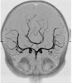
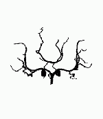
First the anatomical 3D images are processed and segmented in order to label the voxels that belong to the desired arteries. This process is illustrated in the next two images where the maximum intensity projection (MIP) of the images before and after segmentation are shown.


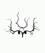
After segmentation a geometrical model is constructed by tessellating the images. This model is then cut in order to simulate only a desired portion of the arterial tree. These processes are illustrated in the following two images.
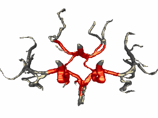
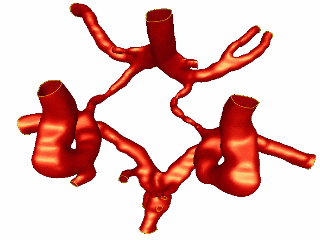
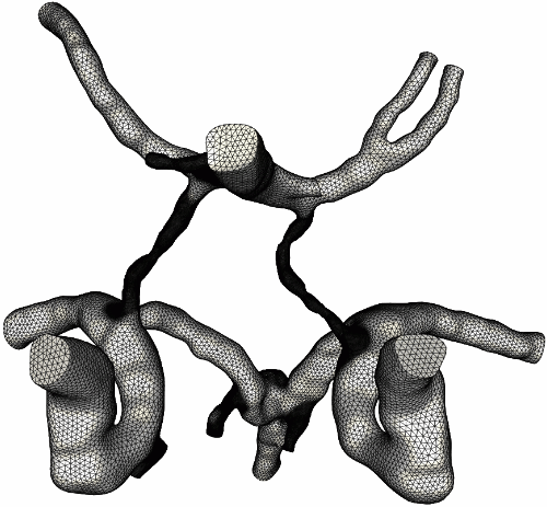
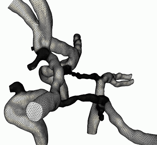
This final model is the used to generate finite element grids for the CFD calculations. The following three images show the surface of the finite element grid from different view-points.

Observations: