Description:
In this case the blood flow in a rigid model of the renal arteries of a normal subject is computed. The anatomical model was reconstructed from contrast-enhanced MRA images and flow conditions were obtained using phase-contrast MRA velocity measurements.
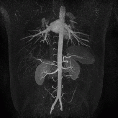
The MIP projection of the contrast-enhanced MRA images is shown below:

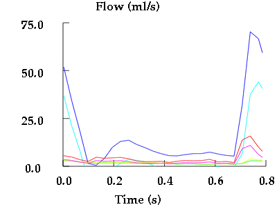
The flow waveforms measured using phase-contrast MRA for different arterial branches (renals, aorta, sma and celiac) are shown below:
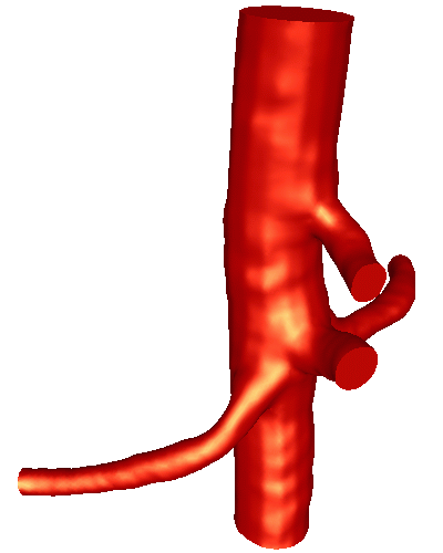
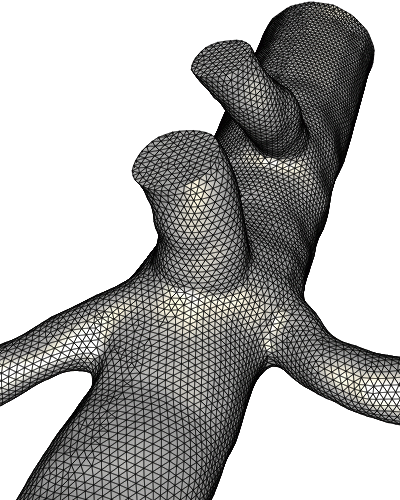
The reconstructed anatomical model and the generated finite element grid are shown below:
The following images show the velocity magnitude on two perpendicular planes at different times during the cardiac cycle:
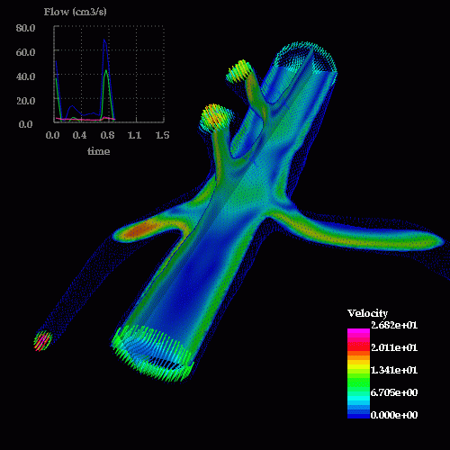
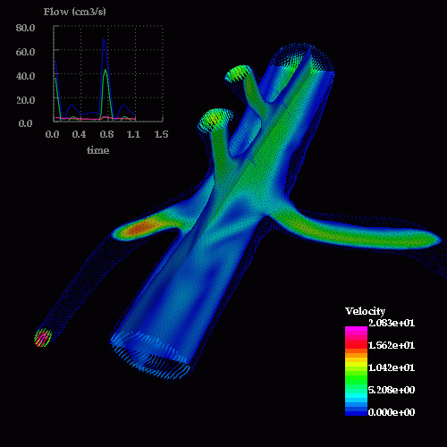
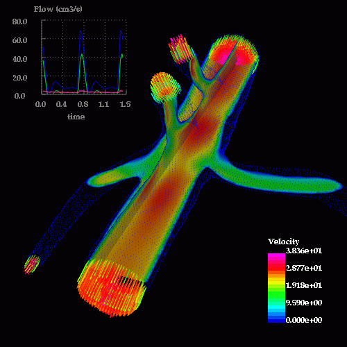
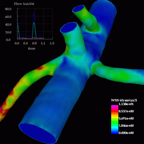
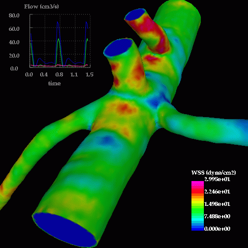
The following images show the wall shear stress distribution at the same times instants during the cardiac cycle:
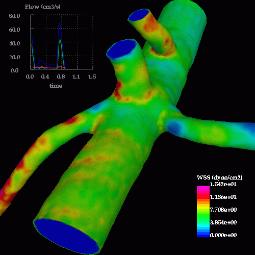
Observations: